Are Lobulated masses cancer
Probably benign masses: BI-RADS 3. In mammography, they are well-circumscribed, round, oval or lobulated masses and are not calcified or liquid in sonography. The vast majority of these masses correspond to benign lesions such as a fibroadenoma or cyst with thick contents.
What does it mean to have a nodule in the breast?
The lumps are milk ducts and tissues around them that have grown and widened to form cysts. These enlarge quickly in response to hormones released near your period. The lumps may be hard or rubbery and could feel like a single (large or small) lump. Fibrocystic changes can also cause breast tissue to thicken.
Can an irregular breast mass be benign?
Irregular hypoechoic masses in the breast do not always indicate malignancies. Many benign breast diseases present with irregular hypoechoic masses that can mimic carcinoma on ultrasonography.
Are breast nodules usually cancerous?
As we have seen, most breast lumps are benign, non-cancerous cysts or tumors. Although they may require surgical removal to prevent their interfering with normal breast function, they will not invade surrounding tissue; they are not life threatening.Why is a biopsy done on the breast?
Breast biopsies may be done: To check a lump or mass that can be felt (is palpable) in the breast. To check a problem seen on a mammogram, such as small calcium deposits in breast tissue (microcalcifications) or a fluid-filled mass (cyst) To evaluate nipple problems, such as a bloody discharge from the nipple.
Can you tell if a mass is cancerous from an ultrasound?
Ultrasound cannot tell whether a tumor is cancer. Its use is also limited in some parts of the body because the sound waves can’t go through air (such as in the lungs) or through bone.
Is a Lobulated breast mass cancer?
Unlike more common breast cancers, lobular breast cancer is less likely to form into a lump in the breast tissue or under the arm. Instead, you may feel a fullness, thickening or swelling in one area that feels different from the surrounding area.
Are breast nodules common?
If you find a breast lump or other change in your breast, you might worry about breast cancer. That’s understandable. But breast lumps are common, and most often they’re noncancerous (benign), particularly in younger women.What does Lobulated hypoechoic mass mean?
A hypoechoic mass is tissue in the body that’s more dense or solid than usual. This term is used to describe what is seen on an ultrasound scan. Ultrasound uses sound waves that are absorbed by or bounce off of tissues, organs, and muscles.
What does nodule mean on mammogram?A potential abnormality on a mammogram might be called a nodule, mass, lump, density, or distortion: A mass (lump) with a smooth, well-defined border is often benign. Ultrasound is needed to see and describe the inside of a mass. If the mass contains fluid, it is called a cyst.
Article first time published onWhat does a nodule in the breast look like?
A round, smooth and firm breast lump. A large, solid-feeling lump that moves easily under your skin. A hard, irregular-shaped breast lump. Skin redness or dimpling like an orange.
What is in a nodule?
A nodule is a growth of abnormal tissue. Nodules can develop just below the skin. They can also develop in deeper skin tissues or internal organs. Dermatologists use nodules as a general term to describe any lump underneath the skin that’s at least 1 centimeter in size.
Do nodules in the breast go away?
It’s hard not to panic when you discover a breast lump. Fortunately, most lumps aren’t cancerous. Your healthcare provider can order the appropriate tests to determine what’s causing benign breast disease. Most people don’t need treatment — lumps go away on their own.
What quadrant has most breast cancers?
Most breast cancers develop in the upper outer quadrant of the breast, closest to the armpit. This is because this area has a lot of glandular tissue.
What does echoic mean in ultrasound?
Echogenicity (misspelled sometimes as echogenecity) or echogeneity is the ability to bounce an echo, e.g. return the signal in ultrasound examinations. In other words, echogenicity is higher when the surface bouncing the sound echo reflects increased sound waves.
What is a hypoechoic nodule in the breast?
Hypoechoic nodule or solid lesion in a breast Hypoechoic means an area looks darker on ultrasound than the surrounding tissue. The surrounding tissue therefore looks brighter/lighter shades of grey.
Why would I need an ultrasound after a mammogram?
Why might I need a breast ultrasound? A breast ultrasound is most often done to find out if a problem found by a mammogram or physical exam of the breast may be a cyst filled with fluid or a solid tumor. Breast ultrasound is not usually done to screen for breast cancer.
How painful is a breast biopsy?
You will be awake during your biopsy and should have little discomfort. Many women report little pain and no scarring on the breast. However, certain patients, including those with dense breast tissue or abnormalities near the chest wall or behind the nipple, may be more sensitive during the procedure.
Should you wear a bra after a breast biopsy?
Wear a tight-fitting bra to help support your biopsy site and make you feel more comfortable. Your radiologist will let you know if you need to wear any special type of bra after your biopsy. For 3 days after your biopsy, do not: Lift anything heavier than 5 pounds (2.3 kilograms).
How long do you have to wear a bra after breast biopsy?
The bra will provide comfort and support after your procedure. Please wear a bra for three to four days following surgery, even while you sleep. This minimizes post-operative bleeding and will make you more comfortable.
What does lobular mean in medical terms?
Medical Definition of lobular : of, relating to, affecting, or resembling a lobule lobular fatty degeneration of the liver— Leopold Bellak. Other Words from lobular.
What does Lcis mean in breast cancer?
Lobular carcinoma in situ (LCIS) is a type of breast change that is sometimes seen when a breast biopsy is done. In LCIS, cells that look like cancer cells are growing in the lining of the milk-producing glands of the breast (called the lobules), but they don’t invade through the wall of the lobules.
How fast does lobular breast cancer grow?
According to the Robert W. Franz Cancer Research Center at Providence Portland Medical Center, breast cancer cells need to divide at least 30 times before they are detectable by physical exam. Each division takes about 1 to 2 months, so a detectable tumor has likely been growing in the body for 2 to 5 years.
What is the next step after a breast ultrasound?
The most likely next step is a diagnostic mammogram or breast ultrasound. In some cases, a breast MRI or a biopsy may be recommended. Here are the different types of follow-up tests: Mammography can be used as a follow-up test when something abnormal is found on a screening mammogram or CBE.
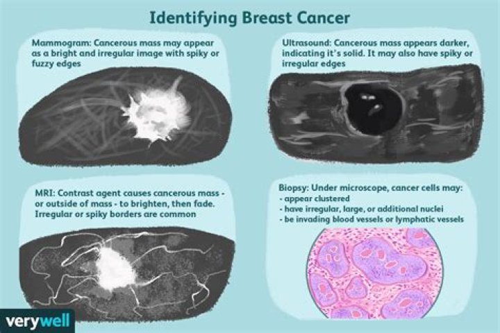
What does a cancerous breast lump look like on an ultrasound?
On ultrasound, a breast cancer tumor is often seen as hypoechoic. It has irregular borders, and may appear spiculated. Other ultrasound findings that suggest breast cancer include: Nonparallel orientation (not parallel to the skin)
Which is better CT scan or ultrasound?
In terms of procedures, ultrasound is used in prenatal care, removal of gall stones, kidney stones, and many other types of medical applications. In both cases, CT and ultrasound are often preferable over regular x-rays. The CT offers a much better image and it can be directed precisely at a target area.
Can a hypoechoic nodule be benign?
Spongiform nodules, purely or predominantly cystic nodules, nodules with well-defined hypoechoic halo and echogenic as well as isoechoic nodules are usually benign. None of the US characteristics have 100% accuracy in detecting or excluding malignancy.
What percentage of hypoechoic nodules are malignant?
About 2 or 3 in 20 are malignant, or cancerous. Malignant nodules can spread to surrounding tissues and other parts of the body. Solid nodules in your thyroid are more likely to be malignant than fluid-filled nodules, but they’re still rarely cancerous.
Is hypoechoic good or bad?
Solid masses are hypoechoic and can be cancerous. Cysts filled with air or fluid are usually hyperechoic and are rarely cancerous. Abnormal tissue also looks different from healthy tissue on a sonogram. Your doctor will usually do further testing if an ultrasound shows a solid mass or what looks like abnormal tissue.
Is a breast nodule the same as a cyst?
What’s the Difference Between a Breast Cyst and a Tumor? Finding a breast lump can be unsettling, but most breast lumps aren’t cancerous. Very often, these lumps turn out to be fluid-filled cysts. There are some characteristics that can help differentiate a cyst from a tumor.
What kind of breast lump should I worry about?
Lumps that feel harder or different from the rest of the breast (or the other breast) or that feel like a change are a concern and should be checked. This type of lump may be a sign of breast cancer or a benign breast condition (such as a cyst or fibroadenoma).