Is LBBB bifascicular block
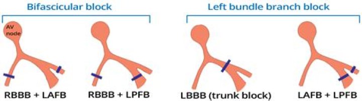
LBBB alone is not considered bifascicular block (LAFB plus LPFB), although anatomically this may be the case. Bifascicular block occurs in 1% to 2% of the adult population.
What are the three fascicles?
A trifascicular block is the combination of a right bundle branch block, left anterior or posterior fascicular block and a first-degree AV block (prolonged PR interval).
What is bifascicular block and trifascicular block?
True trifascicular block refers to the presence of conduction delay in all three fascicles below the AV node (RBBB, LAFB, LPFB), manifesting as bifascicular block and 3rd degree AV block. One of two ECG patterns is present: 3rd degree AV block + RBBB + LAFB or; 3rd degree AV block + RBBB + LPFB.
How can you tell if your block is Bifascicular?
- Conduction to the ventricles is via the single remaining fascicle.
- The ECG will show typical features of RBBB plus either left or right axis deviation.
- RBBB + LAFB is the most common of the two patterns.
How long can I live with Bifascicular block?
Results: During a median follow-up of 84 months, 33 patients died, of whom 14 in SCD. In a univariate analysis, high age, a previous myocardial infarction, and CHF were associated with a significantly increased risk of all-cause mortality and SCD.
What's Fascicular block?
Fascicular block involves the anterior or posterior fascicle of the left bundle branch. Interruption of the left anterior fascicle causes left anterior hemiblock characterized by modest QRS prolongation (< 120 millisecond) and a frontal plane QRS axis more negative than −30° (left axis deviation).
What is the ICD 10 code for Bifascicular block?
I45. 2 is a billable/specific ICD-10-CM code that can be used to indicate a diagnosis for reimbursement purposes.
Which is more serious LBBB or RBBB?
Conclusions. In patients with LVEF<35%, RBBB is associated with significantly greater scar size than LBBB and occlusion of a proximal LAD septal perforator causes RBBB. In contrast, LBBB is most commonly caused by nonischemic pathologies.Is bifascicular block life threatening?
Patients with bifascicular block (BFB) have a high mortality rate. The purpose of the present study was to identify high-risk patients in a BFB population by performing an extensive cardiac evaluation including noninvasive and invasive tests.
How do you treat bifascicular block?Treatment. In those with bifascicular block and no symptoms, little with respect to treatment is needed. In those with syncope, a pacemaker is recommended.
Article first time published onCan bifascicular block cause syncope?
Syncope is a common event in bifascicular block patients and its causes are difficult to assess. However, results from the PRESS study have found that by using a dual chamber pacemaker programmed to 60ppm lower rate the number of syncope events can be reduced.
Is LAFB life threatening?
The condition, called “left anterior fascicular block” (LAFB), involves scarring in a section of the hearts’ left ventricle (pumping chamber). People with the condition may be at higher risk of heart failure, sudden cardiac death or a dangerous heart rhythm disorder called atrial fibrillation, the new study found.
Can you have both Rbbb and LBBB?
Alternating Bundle Branch Block (ABBB) is when both right bundle branch block (RBBB) and left bundle branch block (LBBB) patterns appear on the same ECG or within a period of hours to days [1].
What is AV node block?
AV block is a term to describe abnormal impulse conduction through the AV node. This can be manifest as slower than usual propagation through the AV nodal structure to the ventricles, or complete disconnection of the atrial electrical signals to the ventricles.
What causes 3rd degree heart block?
Third-degree heart block may be caused by: Damage to the heart from surgery. Damage to the heart muscle from a heart attack. Other types of heart disease that result in heart muscle damage.
Can Rbbb cause sudden death?
A pattern of RBBB with right axis deviation (RAD) increased the risk of sudden death during follow-up. Four of 14 patients with RBBB RAD died suddenly during follow-up.
What is considered structural heart disease?
“Structural heart disease is a form of heart disease that refers to defects within your heart that you were either born with or have developed due to aging, injury, or infection. Similar to other kinds of heart disease, it can lead to health problems if left untreated.
Is LBBB hereditary?
Hereditary bundle branch defect is an autosomal dominant genetic disease that, in a large Lebanese family, was mapped to the long arm of chromosome 19.
What is medical term LAFB?
Left anterior fascicular block (LAFB) is considered a failure or delay of conduction in the left anterior fascicle. 1. Despite the fact that little is known about the long-term prognosis associated with LAFB, it has generally been thought of as a benign electrocardiographic (ECG) finding.
What is the ICD 10 code for sick sinus syndrome?
I49. 5 is a billable/specific ICD-10-CM code that can be used to indicate a diagnosis for reimbursement purposes.
What is the ICD-10-CM code for prinzmetal angina?
Prinzmetal angina and variant angina are coded as angina pectoris with documented spasm, code I20. 1 in ICD-10-CM.
Is Fascicular block serious?
A cardiac condition called left anterior fascicular block (LAFB), in which scarring occurs in a section of the left ventricle, may not be as benign as currently thought and could increase the likelihood of heart failure, sudden cardiac death or atrial fibrillation.
Is left anterior fascicular block the same as LBBB?
Left anterior fascicular block (LAFB) is an abnormal condition of the left ventricle of the heart, related to, but distinguished from, left bundle branch block (LBBB). It is caused by only the anterior half of the left bundle branch being defective. It is manifested on the ECG by left axis deviation.
Can you live a normal life with left bundle branch block?
In young and healthy people, left bundle branch block is rare. This condition seems to have little effect on how long you live if you have no other underlying heart problems. You may not need any treatment at all, . especially when you have no other disease affecting your heart.
What is RBB in ECG?
Right bundle branch block (RBBB) is an abnormal pattern that is seen on the electrocardiogram (ECG), which indicates that the heart’s electrical impulse is not being distributed normally across the ventricles.
What is Echo complete?
An echocardiogram checks how your heart’s chambers and valves are pumping blood through your heart. An echocardiogram uses electrodes to check your heart rhythm and ultrasound technology to see how blood moves through your heart.
Is Sinus a rhythm?
Sinus rhythm is the name given to the normal rhythm of the heart where electrical stimuli are initiated in the SA node, and are then conducted through the AV node and bundle of His, bundle branches and Purkinje fibres. Depolarisation and repolarisation of the atria and ventricles show up as 3 distinct waves on ECG.
What is LVH?
Left ventricular hypertrophy is a thickening of the wall of the heart’s main pumping chamber. This thickening may result in elevation of pressure within the heart and sometimes poor pumping action. The most common cause is high blood pressure.
Is exercise good for LBBB?
After three months of regular exercise training with 30-minute sessions per day for five days a week, the patient’s symptoms improved with development of LBBB and chest pain at a considerably higher heart rate of 150 bpm (Figure 3). The morphology of the LBBB remained the same.
Is syncope the same as fainting?
Syncope (SINK-a-pee) is another word for fainting or passing out. Someone is considered to have syncope if they become unconscious and go limp, then soon recover. For most people, syncope occurs once in a great while, if ever, and is not a sign of serious illness.
What is the number one cause of syncopal episodes?
Vasovagal syncope is the most common type of syncope. It is caused by a sudden drop in blood pressure, which causes a drop in blood flow to the brain. When you stand up, gravity causes blood to settle in the lower part of your body, below your diaphragm.