Is the vomer a cranial bone
Located in the center of the nasal cavity, the vomer is a thin, unpaired bone of the face and skull (cranium). This small, trapezoidal bone serves as part of the nasal septum, which is the middle wall of the nasal respiratory cavity.
What type of bone is the vomer?
1. Flat Bones Protect Internal Organs. There are flat bones in the skull (occipital, parietal, frontal, nasal, lacrimal, and vomer), the thoracic cage (sternum and ribs), and the pelvis (ilium, ischium, and pubis). The function of flat bones is to protect internal organs such as the brain, heart, and pelvic organs.
What is the vomer a part of?
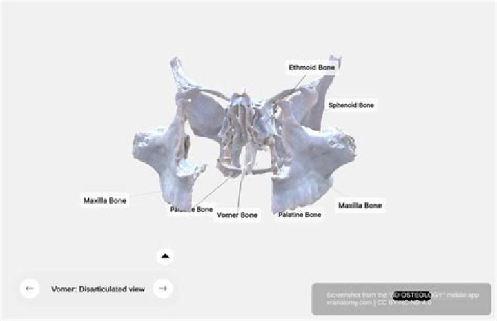
The vomer22 (Fig. 5-58) is a thin, trapezoid-shaped plate of bone that lies in the midline and forms part of the nasal septum. It articulates with the sphenoid, ethmoid and palatine bones, and with the maxilla and septal cartilage (Figs.
What are the cranial bones?
- Parietal (2)
- Temporal (2)
- Frontal (1)
- Occipital (1)
- Ethmoid (1)
- Sphenoid (1)
What are the cranial and facial bones?
The cranium (skull) is the skeletal structure of the head that supports the face and protects the brain. … The facial bones underlie the facial structures, form the nasal cavity, enclose the eyeballs, and support the teeth of the upper and lower jaws.
Which of the following bones does the vomer articulate?
The vomer articulates with the palatine, maxilla, ethmoid and sphenoid bones.
Is the vomer a facial bone?
Located in the center of the nasal cavity, the vomer is a thin, unpaired bone of the face and skull (cranium). This small, trapezoidal bone serves as part of the nasal septum, which is the middle wall of the nasal respiratory cavity.
What are 8 cranial bones?
- Frontal bone. This is the flat bone that makes up your forehead. …
- Parietal bones. This a pair of flat bones located on either side of your head, behind the frontal bone.
- Temporal bones. …
- Occipital bone. …
- Sphenoid bone. …
- Ethmoid bone.
Where is vomer bone?
The vomer is a small, thin, plow-shaped, midline bone that occupies and divides the nasal cavity. It articulates inferiorly on the midline with the maxillae and the palatines, superiorly with the sphenoid via its wings, and anterosuperiorly with the ethmoid.
Where is the cranial?The cranium—the part of the skull that encloses the brain—is sometimes called the braincase, but its intimate relation to the sense organs for sight, sound, smell, and taste and to other structures makes such a designation somewhat misleading.
Article first time published onHow many vomer bones are there?
Components of viscerocranium : The face is made of 2 nasal, 2 lacrimal, 2 palantine, 2 inferior nasal concha, 2 zygomatic, 2 maxilla, 1 mandible, and 1 vomer.
Is the vomer part of the axial skeleton?
The 14 facial bones are the nasal bones, the maxillary bones, zygomatic bones, palatine, vomer, lacrimal bones, the inferior nasal conchae, and the mandible. … Although it is not found in the skull, the hyoid bone is considered a component of the axial skeleton.
Does the vomer bone move?
Scratch your ear. Stuffy nose? … Pushing on both places at the same time moves the vomer bone, which runs through the nasal passages, back and forth like a seesaw. The motion loosens the congestion and after about 20 seconds, the sinuses begin to drain.
What are the 22 cranial and facial bones?
The skull (22 bones) is divisible into two parts: (1) the cranium, which lodges and protects the brain, consists of eight bones (Occipital, Two Parietals, Frontal, Two Temporals, Sphenoidal, Ethmoidal) and the skeleton of the face, of fourteen (Two Nasals, Two Maxillae, Two Lacrimals, Two Zygomatics, Two Palatines, Two …
What are the 6 facial bones?
The primary bones of the face are the mandible, maxilla, frontal bone, nasal bones, and zygoma. Facial bone anatomy is complex, yet elegant, in its suitability to serve a multitude of functions.
What are the 14 facial bone?
The names of the 14 facial bones are: inferior nasal concha (2 of them,) lacrimal bones (2), mandible, maxilla (2), nasal bones (2), palatine bones (2), vomer, and zygomatic bones, or zygoma (2).
Is the vomer the septum?
The vomer22 (Fig. 5.58) is a thin, trapezoid-shaped plate of bone that lies in the midline and forms part of the nasal septum. It articulates with the sphenoid, ethmoid, and palatine bones, and with the maxilla and septal cartilage (Figs.
Is the vomer paired or unpaired?
The paired bones are the maxilla, palatine, zygomatic, nasal, lacrimal, and inferior nasal conchae bones. The unpaired bones are the vomer and mandible bones.
Does the vomer forms part of the hard palate?
The maxilla articulates with both the nasal bone and the vomer. Identify the bone(s) that form(s) the majority of the hard palate and a keystone bone of the face. These bones, along with the palatine bones, form the hard palate. … The vomer forms the inferior portion of the septum.
Is the vomer part of the ethmoid bone?
The vomer forms the inferior part of the nasal septum, with the superior part formed by the perpendicular plate of the ethmoid bone. The name is derived from the Latin word for a ploughshare and the shape of the bone.
Why are the maxillae considered the keystone bones of the face?
Explanation: It is the keystone because it anchors all facial bones except the mandible. It consists of two bones (maxillae) fused together at the midline. Each maxilla forms joints with seven other facial bones plus two of the cranium.
What bone does not articulate with the frontal bone?
The single frontal bone forms the anterior portion of the cranium, anterior floor of the cranial cavity, and superior part of the face (Figure 8-4). At the top of the skull, the frontal bone articulates with the parietal bones; inferiorly it articulates with the sphenoid bone, ethmoid bone, and lacrimal bones.
Which is not a cranial bone?
Which bone is NOT considered to be part of the cranium? The lacrimal bone is a tiny bone found in the medial portion of the orbit. It is a facial bone, not part of the cranium.
Which bone has a prominent head?
Occipital boneHuman skull (Occipital bone is at bottom right).Position of occipital bone (shown in green)DetailsArticulationsthe two parietals, the two temporals, the sphenoid, and the atlas
What is the keystone bone of the face?
Since it anchors all facial bones except the mandible, the maxilla is known as the “keystone bone” of the face. It is made up of two bones (maxillae) that are fused together in the middle. Seven other facial bones, as well as two cranial bones, form joints in each maxilla.
Are there two parietal bones?
The two parietal bones articulate at the sagittal borders with each other to form the sagittal suture. … The squamosal border comes in contact with three bony structures. From anterior to posterior these are the greater wing of the sphenoid bone and the squamous and petrous parts of temporal bone.
What are the 3 bone groups of the skull?
- Parietal Bone. The parietal bone forms most of the upper lateral side of the skull (see [link]). …
- Temporal Bone. …
- Frontal Bone. …
- Occipital Bone. …
- Sphenoid Bone. …
- Ethmoid Bone. …
- Maxillary Bone. …
- Palatine Bone.
What are the 80 bones of the axial skeleton?
- The skull, which contains 22 bones, from which 8 are cranial and 14 are facial,
- 6 middle ear ossicles (3 in each ear),
- 1 hyoid bone in the neck,
- 26 bones of vertebral column,
- 1 chest bone (sternum), and.
- 24 ribs (12 pairs).
What is back of head called?
The occipital bone is a bone that covers the back of your head; an area called the occiput. The occipital bone is the only bone in your head that connects with your cervical spine (neck). The occipital bone surrounds a large opening known as the foramen magnum.
Where is the lacrimal bones?
The lacrimal bones are small, flat craniofacial bones located in the eye socket. These rectangular bones consist of two surfaces, one facing the nose, the other facing the eye.
Where are the femur?
femur, also called thighbone, upper bone of the leg or hind leg. The head forms a ball-and-socket joint with the hip (at the acetabulum), being held in place by a ligament (ligamentum teres femoris) within the socket and by strong surrounding ligaments.