What are myosin components
The main constituent of the thick filaments is myosin. Each thick filament is composed of about 250 molecules of myosin. Myosin has two important roles: a structural one, as the building block for the thick filaments, and a functional one, as the catalyst of…
What are the two sites on a myosin head?
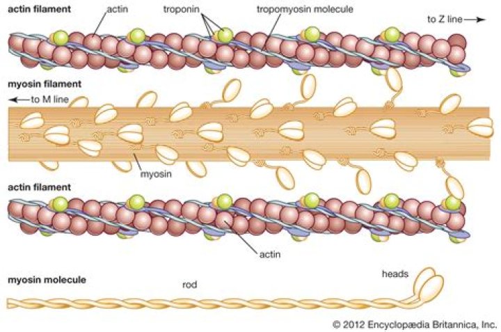
The globular myosin heads extend outward and form cross-bridges when they interact with thin filaments. The myosin heads have two reactive sites: One allows it to bind with the actin filament, and one binds to ATP.
Where are the myosin molecules located?
Where Is Myosin Found? In both eukaryotic cells, cells that have membrane-bound organelles and a nucleus, and prokaryotic cells, cells that lack a nucleus and membrane-bound organelles, we can find myosin. It exists as a filament inside of the cell.
What are the 2 filaments of a muscle?
A sarcomere is the basic contractile unit of muscle fiber. Each sarcomere is composed of two main protein filaments—actin and myosin—which are the active structures responsible for muscular contraction.What proteins make up myosin filaments?
2. Actomyosin ATPase. Myofibrillar proteins include those of the thick filament (mainly myosin) and thin filament (mainly actin, troponin, and tropomyosin). Native mammalian cardiac myosin is composed of two myosin heavy chains (HC) and four myosin light chains (LC).
Why does myosin have two heads?
Several classes of the myosin superfamily are distinguished by their “double-headed” structure, where each head is a molecular motor capable of hydrolyzing ATP and interacting with actin to generate force and motion. … These data suggest that muscle myosins require both heads to generate maximal force and motion.
What are the three parts of myosin?
Structure and functions Most myosin molecules are composed of a head, neck, and tail domain.
What are myosin filaments?
Myosin filaments (also called thick filaments) are key components of muscle and non-muscle cells. In striated muscle, they overlap with thin (actin-containing) filaments in an orderly array, making a repeating pattern of sarcomeres, the basic units of contraction [1] (Figure 1a).What molecules release myosin from actin?
Myosin has another binding site for ATP at which enzymatic activity hydrolyzes ATP to ADP, releasing an inorganic phosphate molecule and energy. ATP binding causes myosin to release actin, allowing actin and myosin to detach from each other.
Where is myosin thick filament located?The thick filament is located at the center of the sarcomere as the giant elastic protein connectin/titin spans half sarcomere along the thick filaments, linking the Z‐band and the M‐lines (Labeit & Kolmerer, 1995; Maruyama, 1976; Wang, McClure, & Tu, 1979).
Article first time published onIs myosin the thick filament?
The thick filament consists largely of myosin. Six proteins make up myosin: two heavy chains whose tails intertwine to form a supercoil and whose heads contain actin binding sites and a catalytic site for ATP hydrolysis.
Which of the following two structures meet to form the neuromuscular junction?
The structures that meet at the neuromuscular junction are (d) the synaptic terminus and the sarcolemma.
What is produced by the two proteins called actin and myosin that are found in the cells of the muscles?
Actin and myosin are both proteins that are found in every type of muscle tissue. Thick myosin filaments and thin actin filaments work together to generate muscle contractions and movement. Myosin is a type of molecular motor and converts chemical energy released from ATP into mechanical energy.
What structure joins myosin filaments in disk A?
The M line region are the sites of titin filaments anchorage which, in the number of 6, twist around the myosin filaments and join with the Z line stabilizing the myosin filaments in the sarcomeres.
How is myosin synthesized?
The steps in its synthesis are; Transcription, where the nucleotide sequence in a myosin gene is copied to form mRNA. mRNA undergoes modification to safely reach ribosomes in the cytoplasm. Translation, during which amino acids are arranged in a sequence according to mRNA to make myosin molecule.
What are the 2 main proteins required for muscles to contract?
Muscles are composed of two major protein filaments: a thick filament composed of the protein myosin and a thin filament composed of the protein actin. Muscle contraction occurs when these filaments slide over one another in a series of repetitive events.
What two proteins regulate the attachment of the myosin head to the actin binding site?
This recombination step between the myosin-products complex and actin is controlled by the regulatory proteins troponin and tropomyosin in response to calcium ion concentrations. The force for contraction is generated by movement of the cross-bridge head to a 45° angle of attachment (Step 5, Fig.
What are the types of myosin?
Three types of unconventional myosins predominate: myosin I, myosin V, and myosin VI. The unconventional myosin I and V categories contain multiple members.
What is myosin quizlet?
myosin definition. contractile protein that is thick located in myofilaments. sarcomere. the contractile unit of muscle extends to Z-line to Z-line.
Does myosin have 2 heads?
Myosin has two heads which can bind with F-actin and react with ATP. The skeletal muscle myosin forms each 1 mol of the myosin-phosphate-ADP complex (M-P-ADP) and the myosin-ATP complex (M-ATP).
What binds to the myosin head?
In addition to binding actin, the myosin heads bind and hydrolyze ATP, which provides the energy to drive filament sliding. This translation of chemical energy to movement is mediated by changes in the shape of myosin resulting from ATP binding.
Why are myosin heads called cross bridges?
Myosin heads are called cross-bridges because they form bridges between the myosin and actin in order to move the actin filaments and cause muscular contraction. The high energy configuration of the actin heads extends the myosin to the actin, reminiscent of a drawbridge.
What change occurs in the myosin molecule between stage 1 and 2?
The change that occurs in the myosin molecule between stage 1 (attachment) and stage 2 (pulling). The myosin head doesn’t release from actin binding site until a new molecule enters the cycle. According to the model, what molecule allows myosin to release from actin?
What molecule is connected to the Z line?
Actin filaments and titin molecules are cross-linked in the Z-disc via the Z-line protein alpha-actinin. The M-band proteins myomesin as well as C-protein crosslink the thick filament system (myosins) and the M-band part of titin (the elastic filaments).
What is released when myosin heads attach to actin filaments?
What is released when myosin heads attach to actin filaments? Explanation: Phosphate is released when myosin heads attaach to actin myofilaments.
Is myosin fibrous or globular?
Myosin is therefore unusual in that it is both a fibrous protein, and a globular enzyme.
Where does myosin get the energy to perform a contraction?
Where does the energy for muscle contraction come from? Adenosine Triphosphate (ATP). How is the energy used in muscle contraction? The myosin head uses the energy from the ATP molecule, causing the ATP to lose a phosphate molecule and become Adenosine Diphosphate (ADP), to detach from the actin.
How many amino acids are in myosin?
In myosin II molecules, there is usually a 26 amino acid separation between the start of IQ motifs, but in unconventional myosins, the separation can be between 23 and 26 residues [32]. For example, in myosin V molecules, the six IQ motifs are separated by an alternating pattern of 23 and 25 residues.
What do thin filaments consist of?
Thin filaments are composed primarily of the contractile protein actin. As illustrated in Figures 2-8, A and B, actin is composed of small globular subunits (G actin) that form long strands called fibrous actin (F actin).
What is the a band?
The A band is the region of a striated muscle sarcomere that contains myosin thick filaments. In fact, the A band is the entire length of the thick filament of the sarcomere. Its length is approximately 1 μm. The center of the A band is located at the center of the sarcomere (M line).
What are the steps of the neuromuscular junction?
- An AP travels down the axon. to the axon terminal.
- Electrical gated calcium channels open. …
- Calcium causes the vesicles to. …
- ACH diffuses across the synaptic cleft. …
- ACH binding opens ion channels. …
- If the muscle reaches the threshold (-55mv) at the motor end plate. …
- ACH is broken down by.