What does a cutaneous horn look like
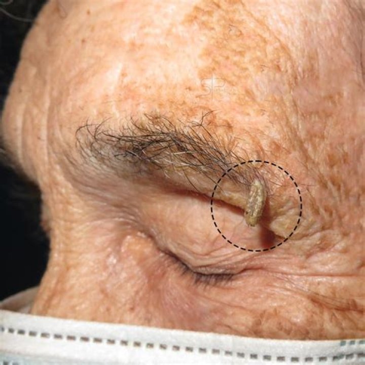
A cutaneous horn looks like a growth on the outside of the skin. This is the most common symptom. It can appear as a large bump, cone, spike, or horn. The growth may be the same color as the skin or it may be a different color.
Is cutaneous horn cancerous?
Even though 60% of the cutaneous horns are benign in nature, the possibility of skin cancer should always be kept in mind. The clinical diagnosis includes various benign and malignant lesions at its base.
How do you treat a cutaneous horn on a dog?
If the lesion that is the underlying cause of a skin horn is benign (noncancerous), it is often treated by excision (surgical removal or resection) or with a procedure called curettage. This is a medical procedure involving the removal of tissue by scraping or scooping.
Is a cutaneous horn a wart?
Cutaneous horn usually arises due to an underlying epidermal lesion, the most common being verruca vulgaris (wart), actinic keratosis (a potentially pre-malignant lesion of dysplastic keratinocytes), or squamous cell carcinoma (a form of skin cancer). These can look essentially identical clinically.How does a cutaneous horn start?
A cutaneous horn, also known as cornu cutaneum, refers to a specific appearance of a skin lesion in which a cone-shaped protuberance arises on the skin caused by overgrowth of the most superficial layer of skin (epidermis). A cutaneous horn is not a particular lesion but is a reaction pattern of the skin.
What percentage of cutaneous horns are cancerous?
Cutaneous horn is a clinical diagnosis observed in both benign and malignant conditions. About 40% of cutaneous horns are part of malignant lesions, especially actinic keratosis, Bowen’s disease and squamous cell carcinoma (Table 1).
Can I cut off a cutaneous horn?
Removing a cutaneous horn The most common treatment for cutaneous horns is removal. The type of treatment you receive will also depend on if the growth is cancerous or noncancerous. Your recovery time will vary depending on the size of the growth and its type.
Do Keratoacanthomas go away?
It’s not unusual for a single keratoacanthoma to shrink and disappear on its own after several months. But it may leave a worse scar than one from surgery. It could also come back, so it’s best to get it removed. If you don’t treat it, keratoacanthoma can spread throughout your body.What are horn cysts?
Horn cysts represent foci of abrupt complete keratinization (with only a very thin surrounding granular cell layer and without retained nuclei). It may be pseudo or true. Pseudo horn cysts results from sectioning of a markedly papillomatous skin.
How do you treat horned paws?To keep the horns from recurring, your vet will excise the base of the growth. Your vet can prescribe Azithromycin or Interferon to reduce pain and discomfort. For the most part, horned paws aren’t something to lose sleep over.
Article first time published onWhat does a cutaneous horn look like on a dog?
A cutaneous horn on a dog will be a growth that sticks up from the skin surface. It can feel like a stick-like growth on a dog’s tail. While they can develop anywhere, they often appear on the back, tail, and legs. They may also ooze pus or blood.
Are cutaneous horns common in dogs?
Cutaneous horns on dogs and cats In cats, especially, cutaneous horns are quite common and usually form on the footpads. The cause is typically feline leukemia virus-associated dermatoses or a feline papillomavirus infection. Less often , cutaneous horns form in dogs as a result of a canine papillomavirus infection.
Can dogs get cutaneous horns?
Cutaneous horns are rare in dogs and cats and causes are speculative, although it is believed that exposure to radiation can trigger the condition. Aetiology comes from an evaluation of the skin at the base or beneath it.
How common is a cutaneous horn?
A cutaneous horn is more common in older patients, with the peak incidence in those between 60 and 70. They are equally common in males and females, although there is a higher risk of the lesion being malignant in men. They are more common in people with fairer skins (skin phototype I and 2).
What does a lesion look like?
Skin lesions are areas of skin that look different from the surrounding area. They are often bumps or patches, and many issues can cause them. The American Society for Dermatologic Surgery describe a skin lesion as an abnormal lump, bump, ulcer, sore, or colored area of the skin.
What is Dermatofibrosis?
Dermatofibromas are small, noncancerous (benign) skin growths that can develop anywhere on the body but most often appear on the lower legs, upper arms or upper back. These nodules are common in adults but are rare in children. They can be pink, gray, red or brown in color and may change color over the years.
What is a horned wart?
The cutaneous horn appears as a funnel-shaped growth that extends from a red base on the skin. It is composed of compacted keratin (the same protein in nails). The size and shape of the growth can vary considerably, but most are a few millimeters in length.
What is a sebaceous horn?
The mysterious sebaceous horn (devil’s horn) is a historically perplexing phenomenon of unknown etiology. It classically occurs on the sun‐exposed areas of the face and hands and consists of a keratin mound with a benign base in most cases and squamous cell carcinoma occurring in about 20% of patients.
What is keratosis on face?
An actinic keratosis (ak-TIN-ik ker-uh-TOE-sis) is a rough, scaly patch on the skin that develops from years of sun exposure. It’s often found on the face, lips, ears, forearms, scalp, neck or back of the hands.
How fast do cutaneous horns grow?
The duration of growth or persistence of GCH has been reported from six weeks to seventy-five years. The largest horn was reported by Michal M et al (2002)[4] had a length of 25 cm. The most common histopathological findings at the base of GCH include squamous cell carcinoma[7,8] and verruca vulgaris.
What is lentigo maligna?
Lentigo maligna is a subtype of melanoma in situ that is characterized by an atypical proliferation of melanocytes within the basal epidermis; lentigo maligna that invades the dermis is termed lentigo maligna melanoma.
What is Bowen's disease?
Bowen’s disease is a very early form of skin cancer that’s easily treatable. The main sign is a red, scaly patch on the skin. It affects the squamous cells, which are in the outermost layer of skin, and is sometimes referred to as squamous cell carcinoma in situ.
Can you pick off a seborrheic keratosis?
Treatment of a seborrheic keratosis isn’t usually needed. Be careful not to rub, scratch or pick at it. This can lead to itching, pain and bleeding.
How can you tell the difference between melanoma and seborrheic keratosis?
The fact that a patient has several lesions with the same or almost the same appearance, is a strong indication of a diagnosis of seborrheic keratoses. Their greasy or verrucous consistency upon palpation distinguishes them from atypical pigmented naevi and malignant melanomas.
What are horn Pseudocysts?
Benign skin tumors They are oval to round, often with a “greasy,” “waxy,” or verrucous appearance. The presence of surface horn pseudocysts, which represent keratin-filled pits, is virtually pathognomonic.
Do Keratoacanthomas metastasize?
Multiple keratoacanthomas It’s a non-melanoma skin cancer that rarely metastasizes, meaning it won’t spread to other areas of the body. But it can still be dangerous and should be treated by a doctor. Many people with one KA lesion may develop more throughout their lifetime.
Do Keratoacanthomas hurt?
Most keratoacanthoma are painless, though some may be itchy. Depending on the site of involvement, keratoacanthoma may interfere with normal function of the affected area.
What does a cancerous pimple look like?
A melanoma pimple will typically present itself as a firm red, brown or skin-colored bump that many doctors may misdiagnose as a pimple or harmless blemish. The main difference to note is that these bumps will not feel soft like a pimple, but rather will be firm or hard to the touch.
How do I get rid of cutaneous horn cat?
The horny growth can be removed by trimming, however, the horns will often recur. Horns causing discomfort should be removed and, if it is possible to do so without causing a large pad defect, the base of the lesion should be excised to prevent re-growth.”
What causes overproduction of keratin in cats?
Contributing factors may include poor grooming habits of the cat, genetic tendency to produce too much sebum (a naturally produced oil produced by the skin glands), improper shedding of the hair thus leading to clogged follicles, or abnormal keratin production.
What causes pillow foot in cats?
Pododermatitis occurs when the immune system is mistakenly triggered and it overproduces lymphocytes that then pool in the cat’s foot pads. Antibodies then attack healthy paws and cause swelling and pain to develop.