What is excitation of the heart
Automatic Electrical Rhythmicity of the Sinus Fibers Some cardiac fibers have the capability of self-excitation, a process that can cause automatic rhythmical discharge and contraction. This is especially true of the fibers of the heart’s specialized conducting system, including the fibers of the sinus node.
What is self excitation of heart?
Automatic Electrical Rhythmicity of the Sinus Fibers Some cardiac fibers have the capability of self-excitation, a process that can cause automatic rhythmical discharge and contraction. This is especially true of the fibers of the heart’s specialized conducting system, including the fibers of the sinus node.
What are the steps of excitation?
The EC-coupling cycle involves the following sequence of events: (1) depolarization of the plasma membrane and its membrane invaginations (the t-tubular system) by an action potential; (2) transduction of the depolarization signal to the sarcoplasmic reticulum (SR) membrane; (3) activation of Ca2+ release from the SR …
How does excitation spread in the heart?
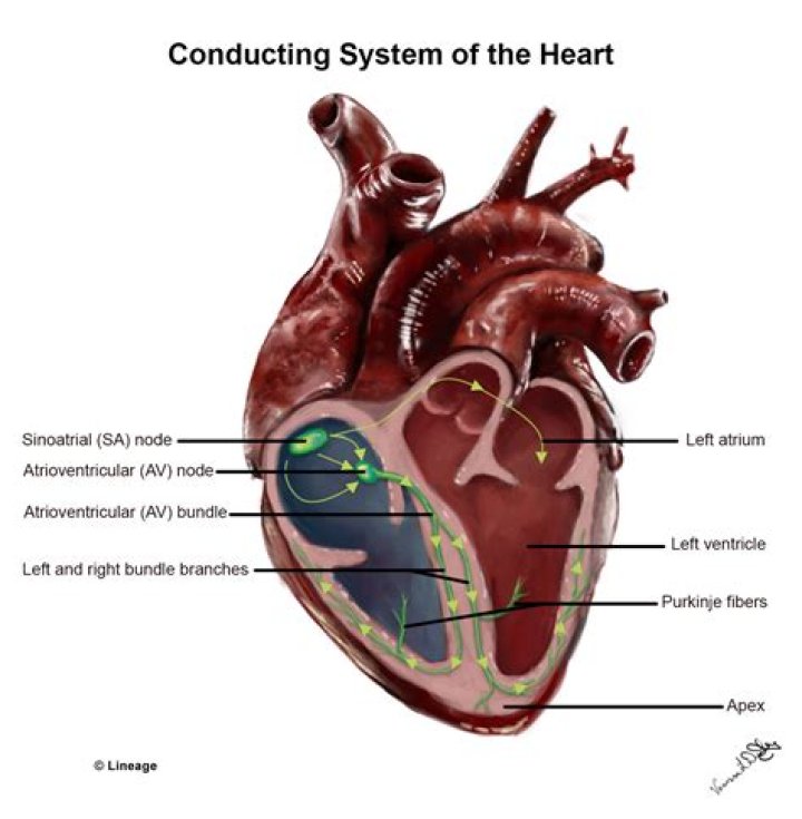
The conduction system of the heart. Left: Normal excitation originates in the sinoatrial (SA) node then propagates through both atria. The atrial depolarization spreads to the atrioventricular (AV) node, and passes through the bundle of His to the bundle branches/Purkinje fibers.What controls the excitation and contraction of the heart?
Cardiac contractility is regulated by changes in intracellular Ca concentration ([Ca2+]i). … Much of the Ca needed for contraction comes from the sarcoplasmic reticulum and is released by the process of calcium-induced calcium release.
What is another name for the SA node?
The SA node, also known as the sinus node, represents a crescent-like shaped cluster of myocytes divided by connective tissue, spreading over a few square millimeters. It is located at the junction of the crista terminalis in the upper wall of the right atrium and the opening of the superior vena cava.
Is the SA node self exciting?
The sinoatrial node is responsible for the spontaneous depolarisation or self excitation. It is the sinoatrial node which determines the normal heart rate of an individual. It depolarises on average 70 to 80 times a minute.
How does the heart's conduction system work?
Heart Conduction System (Cardiac Conduction) The heart conduction system is the network of nodes, cells and signals that controls your heartbeat. Each time your heart beats, electrical signals travel through your heart. … The expansion and contraction control blood flow through your heart and body.What is the wave of excitation?
in physiology, the propagation of electrical activity through tissue, as through nerve or muscle tissue.
What is conduction pathway of the heart?The main parts of the system are the SA node, AV node, bundle of HIS, bundle branches, and Purkinje fibers. … Next, the signal travels to the AV node, through the bundle of HIS, down the bundle branches, and through the Purkinje fibers, causing the ventricles to contract.
Article first time published onWhat structures are involved in excitation?
- This process involves two closely interrelated sets of internal membrane systems, namely the transverse tubules (T-tubules) and sarcoplasmic reticulum. …
- The triad of skeletal muscle is made up of two cisternae of the sarcoplasmic reticulum and one T-tubule.
What is muscle excitation?
Excitation–contraction coupling occurs when depolarization of skeletal muscle cell results in a muscle action potential, which spreads across the cell surface and into the muscle fiber’s network of T-tubules, thereby depolarizing the inner portion of the muscle fiber.
How is excitation of the sarcolemma?
Excitation refers to the propagation of action potentials along the axon of a motor neuron. Excitation of the sarcolemma is coupled or linked to the contraction of a skeletal muscle fiber. … Calcium release from the sarcoplasmic reticulum initiates the contraction.
Where does cardiac excitation normally begin?
Initiation. Located in the wall of the right atrium is a group of specialised cells, called the Sinoatrial node (SAN). These cells, unlike most other cells within the heart, can spontaneously produce action potentials.
Where does excitation-contraction coupling occur?
First coined by Alexander Sandow in 1952, the term excitation-contraction coupling (ECC) describes the rapid communication between electrical events occurring in the plasma membrane of skeletal muscle fibres and Ca2+ release from the SR, which leads to contraction.
What is the process of excitation-contraction coupling in heart muscle cells?
Excitation–contraction coupling describes the processes relating to electrical excitation through force generation and contraction in the heart. It occurs at multiple levels from the whole heart, to single myocytes and down to the sarcomere.
Why SA node is called pacemaker?
The cells of the SA node at the top of the heart are known as the pacemaker of the heart because the rate at which these cells send out electrical signals determines the rate at which the entire heart beats (heart rate). The normal heart rate at rest ranges between 60 and 100 beats per minute.
Does hyperpolarization cause action potential?
Hyperpolarization is a change in a cell’s membrane potential that makes it more negative. It is the opposite of a depolarization. It inhibits action potentials by increasing the stimulus required to move the membrane potential to the action potential threshold.
Is the SA node a pacemaker?
The SA node is sometimes called the heart’s “natural pacemaker.” The SA node sends electrical impulses at a certain rate, but your heart rate may still change depending on physical demands, stress, or hormonal factors.
Which organ is known as heart of heart?
The heart is a muscular organ about the size of a fist, located just behind and slightly left of the breastbone. The heart pumps blood through the network of arteries and veins called the cardiovascular system.
What is pacemaker of heart?
Overview. A pacemaker is a small device that’s placed (implanted) in the chest to help control the heartbeat. It’s used to prevent the heart from beating too slowly. Implanting a pacemaker in the chest requires a surgical procedure.
What is sinus node heart?
The sinus node is an area of specialized cells in the upper right chamber of the heart. This area controls your heartbeat. Normally, the sinus node creates a steady pace of electrical impulses. The pace changes depending on your activity, emotions, rest and other factors.
Whats is ECG?
An electrocardiogram (ECG or EKG) records the electrical signal from your heart to check for different heart conditions. Electrodes are placed on your chest to record your heart’s electrical signals, which cause your heart to beat. The signals are shown as waves on an attached computer monitor or printer.
How is arrhythmia diagnosed?
An electrocardiogram (EKG or ECG) is the main test for detecting arrhythmia. An EKG records the heart’s electrical activity. Your doctor may do the test while you are at rest or may do a stress test, which records the heart’s activity when it is working hard.
What will happen if an ectopic pacemaker controls the conduction of the heart?
When an ectopic pacemaker initiates a beat, premature contraction occurs. A premature contraction will not follow the normal signal transduction pathway, and can render the heart refractory or incapable of transmitting the normal signal from the SA node.
What is the normal electrical conduction of the heart?
In an adult, the sinus node sends out a regular electrical pulse 60 to 100 times per minute. This electrical pulse travels down through the conduction pathways and causes the heart’s lower chambers (ventricles) to contract and pump out blood.
What are the 3 crucial parts of the cardiac conduction system?
What are the three crucial parts of the cardiac conduction system? Sinoatrial (SA) node, atriaoventricular (AV) node, his-purkinje system. a.k.a. the pace maker; This is a small bundle of cells capable of starting the electrical impulse that will cause the heart to beat.
What triggers the excitation process?
In skeletal muscle fibers, electrochemical activity triggers myofilament movement. These linked events are referred to as excitation-contraction coupling. acetylcholine (ACh) is released by a motor neuron at the neuromuscular junction. action potentials (impulses) that spread out across the sarcolemma.
What chemical that enters a muscle cell upon excitation?
A nerve action potential that is initiated in the cell body of a spinal motor neuron propagates out the ventral roots and eventually invades the synaptic terminals of the motor neurons. As a result of the action potential, the chemical transmitter acetylcholine (ACh) is released into the synaptic cleft.
What happens when troponin and tropomyosin block?
What happens when troponin and tropomyosin block the active sites of actin? The return of calcium ions to the sarcoplasmic reticulum during muscle relaxation decreases the calcium ion concentration in the cytosol.
What stops muscle excitation?
Muscle contraction usually stops when signaling from the motor neuron ends, which repolarizes the sarcolemma and T-tubules, and closes the calcium channels in the SR. Ca++ ions are then pumped back into the SR, which causes the tropomyosin to re-cover the binding sites on actin (Figure 10.3. 2).