What is normal RV TLC ratio
Residual volume (RV) – volume of air in the lungs after a full expiration. RV / TLC ratio : elevated ratio implies air trapping
What should a RV TLC be?
For RV%TLC ratio, the AOC=0.77, sensitivity=77.3 (95%CI – 75.8 to 78.71) and specificity=53.6 (95%CI 51.9 to 55.3). Conclusion: The FRC%TLC is raised in 7% of patients without airflow obstruction, as is the RV%TLC. The worse the airflow obstruction, the greater the ratio observed.
What does a high RV TLC mean?
Elevated RV and RV/TLC ratio suggest air trapping with obstructive lung disease. 4. If available, compare TLC as measured by plethysmography with that measured by gas dilution techniques (such as helium dilution or nitrogen washout).
What does RV TLC ratio tell you?
Increased residual volume and RV/TLC ratio indicate unresolved obstruction in small airways associated with inflammation. Lung volume measurement may be a better tool to assess asthma severity.What does low RV TLC mean?
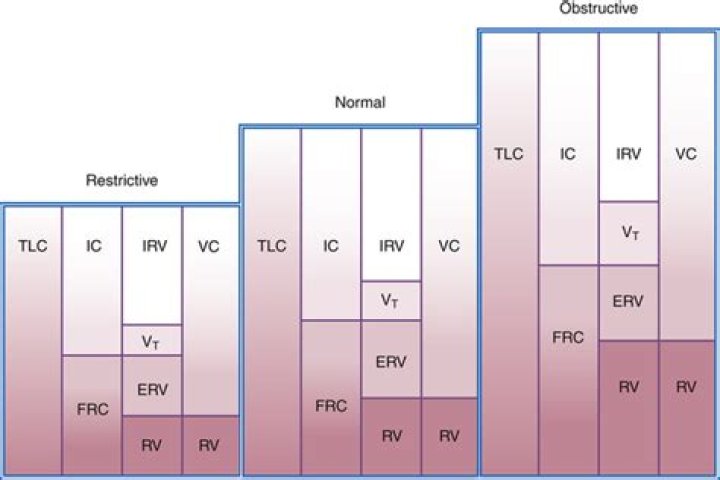
Lung volumes can confirm the presence of restriction when a reduced vital capacity is seen on spirometry. A reduced TLC is the hallmark of restrictive lung disease. An isolated reduction of the residual volume may be an early sign of restrictive lung disease.
How is RV TLC measured?
Once the FRC gas volume is measured and the RV is determined, the following additional equations that can be used to calculate the TLC; the sum of the four lung volumes: TLC = RV + ERV + IRV + TV or the sum of vital capacity and the residual volume: TLC = VC + RV.
What is the normal range for PFT?
Pulmonary function testNormal value (95 percent confidence interval)TLC80% to 120%FRC75% to 120%RV75% to 120%DLCO> 60% to < 120%
What is the normal range for DLCO VA?
Normal DLCO: >75% of predicted, up to 140% Mild: 60% to LLN (lower limit of normal) Moderate: 40% to 60% Severe: <40%Does TLC increase in COPD?
COPD does not generally increase TLC. It just increases residual volume after maximum exhalation. Similarly, increased total lung capacity in obstructive airway defect is primarily caused by increased residual volume.
What is normal residual volume?Residual Volume(RV) It is the volume of air remaining in the lungs after maximal exhalation. Normal adult value is averaged at 1200ml(20‐25 ml/kg) . It is indirectly measured from summation of FRC and ERV and cannot be measured by spirometry.
Article first time published onWhy is RV increased in asthma?
The progressive increase in RV/TLC ratio in patients with longer duration of asthma may be related to chronic inflammatory changes affecting small airway leading to airway closure [17], [19], [20] even in those patients taking inhaled steroids to control asthma symptoms [19], [21], [22], [23].
What does it mean when your lungs are expanded?
Hyperinflated lungs are larger-than-normal lungs as a result of trapped air. It happens when you can’t exhale, or push out all of the air that’s in your lungs. The air gets trapped and takes up space, which can make it harder to get fresh air into your body.
Is DLCO normal in asthma?
Average DLCO values were normal in the asthma group (103%P) and lower in the COPD (69%).
How do you interpret spirometry results?
- 80% or more – mild COPD (able to achieve normal results after medication)
- 50-79% – moderate COPD.
- 30-49% – severe COPD.
- less than 30% – very severe COPD.
Is vital capacity decreased in COPD?
In those with COPD, the airways tend to collapse during a forced expiration due to the reduction of alveolar attachments and airway abnormalities. Therefore, finding that VC is higher than FVC suggests small airway collapse and air trapping [17].
How do you read a respiratory function test?
You will also see another number on the spirometry test results — the FEV1/ FVC ratio. This number represents the percent of the lung size (FVC) that can be exhaled in one second. For example, if the FEV1 is 4 and the FVC is 5, then the FEV1/ FVC ratio would be 4/5 or 80%.
What is abnormal PFT?
Abnormal results usually mean that you may have chest or lung disease. Some lung diseases (such as emphysema, asthma, chronic bronchitis, and infections) can make the lungs contain too much air and take longer to empty.
What is a good lung function test result?
Lung volume is measured in litres. Your predicted total lung capacity (TLC) is based on your age, height, sex and ethnicity, so results will differ from person to person. Normal results typically range between 80% and 120% of the prediction.
How many ml should I be able to inhale?
Respiratory (lung) volumes: Inspiratory reserve volume (IRV) is the amount of air that can be forcibly inhaled beyond a tidal inhalation (about 3,000 ml for men & 2,000 ml for women).
How many Litres is lung capacity?
Did you know that the maximum amount of air your lungs can hold—your total lung capacity—is about 6 liters? That is about three large soda bottles. Your lungs mature by the time you are about 20-25 years old.
How is TLC calculated?
8 Total lung capacity. The total lung capacity (TLC) is the volume of gas in the lung at the end of a full inspiration. It is either calculated from: TLC = RV+IVC, or from: TLC = FRC+IC; the latter is the preferred method in body plethysmography. It can also be measured directly by the radiologic technique.
Can anxiety cause hyperinflation of lungs?
Whether it is exertion, anxiety, agitation, or respiratory distress, any increase in breathing frequency in the setting of airway resistance and expiratory flow limitation can result in dynamic hyperinflation.
What causes decreased lung volumes?
Restrictive lung disease, a decrease in the total volume of air that the lungs are able to hold, is often due to a decrease in the elasticity of the lungs themselves or caused by a problem related to the expansion of the chest wall during inhalation.
When 1200 mL air is left in the lungs it is called?
Residual Volume (RV): Volume of air remaining in the lungs even after a forcible expiration. This averages 1100 mL to 1200 mL.
What does low DLCO indicate?
A reduced DLCO and a reduced KCO suggest a true interstitial disease such as pulmonary fibrosis or pulmonary vascular disease. It has demonstrated that in healthy patients, the KCO is increased to above normal levels when the DLCO test is performed at volumes less than the TLC.
What is low diffusing capacity?
Meaning of a Low Diffusing Capacity Diffusing capacity may be low if lung disease is present that causes the membrane to be thicker, for example, in diseases such as pulmonary fibrosis and sarcoidosis.
What does a low DLCO mean?
Dlco is a specific but insensitive predictor of abnormal gas exchange during exercise. Low Dlco less than or equal to 50% predicted can predict hypoxemia with exercise. A normal Dlco does not rule out oxygen desaturation with exercise.
What is normal residual for G tube feeding?
Typically, standard nursing practice is to stop tube feedings due to gastric residual volume (GRV) that is twice the flow rate. So, a feeding rate of only 40 mL per hour would be held with a measured GRV of 80 mL.
How residual volume is measured?
Residual volume is measured by: A gas dilution test. A person breathes from a container containing a documented amount of a gas (either 100% oxygen or a certain amount of helium in air). The test measures how the concentration of the gases in the container changes.
What causes high residual volume?
Residual volume is the only lung volume that is not decreased with respiratory muscle weakness. Residual volume is the amount of air left in the lungs at the end of a maximal expiration and is typically increased due to the inability to forcibly expire and remove air from the lungs.
Does residual volume decrease in asthma?
Paradoxically the most easily measu- red volume, the vital capacity (VC), is the only volume that decreases as asthma becomes more severe; residual volume (RV) and functional residual capacity (FRC) invariably increase and, more surprisingly, total lung ca- pacity (TLC) itself may increase in the asthmatic attack.