What is the gray commissure
a bundle of nerve fibers that surrounds the central canal of the spinal cord
What does the gray commissure contain?
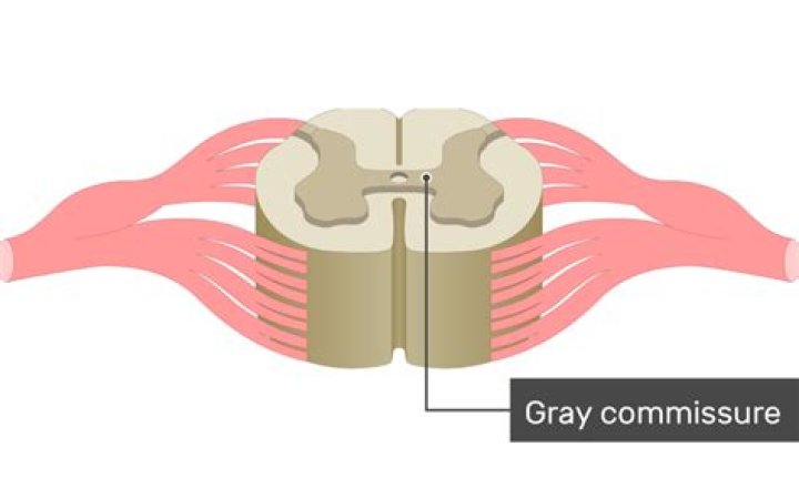
The term gray commissure refers to the bridge of gray matter that contains the central canal of the spinal cord and connects the gray columns on either side of the spinal cord ( Carpenter-1983 ).
What is the difference between gray matter and gray commissure?
Grey matter refers to unmyelinated neurons and other cells of the central nervous system. … The grey matter on the left and right side is connected by the grey commissure. The grey matter in the spinal cord consists of interneurons, as well as the cell bodies of projection neurons.
What is the gray commissure of the spinal cord quizlet?
What is gray commissure of spinal cord? A band of gray matter connecting the gray matter of the left side with the gray matter of the right side. The anterior horns of the spinal cord contain the somas of which neurons? Somatic motor.What is white commissure?
The anterior or ventral white commissure is a collection of nerve fibers that cross the midline of the spinal cord and transmit information from or to the contralateral side of the brain. It is present throughout the length of the spinal cord and lies behind the anterior median fissure.
What does white matter do in the brain?
White matter is tissue in the brain composed of nerve fibers. The fibers (called axons) connect nerve cells and are covered by myelin (a type of fat). The myelin is what gives white matter its white color. Myelin speeds up the signals between the cells, enabling the brain cells to quickly send and receive messages.
What is the cauda?
Cauda is Latin for tail, and equina is Latin for horse (ie, the “horse’s tail”). The CE provides sensory innervation to the saddle area, motor innervation to the sphincters, and parasympathetic innervation to the bladder and lower bowel (ie, from the left splenic flexure to the rectum).
Which of the following is found in the GREY matter of the spinal cord?
The grey matter of the spinal cord contains neuronal cell bodies, dendrites, axons, and nerve synapses.What is the opening between the gray commissure that contains cerebrospinal fluid?
Central canal—opening in the middle of the gray commissure which runs the length of the spinal cord and contains cerebrospinal fluid. Cerebrospinal fluid—fluid which surrounds and cushions the brain and spinal cord, produced in the choroid plexuses within the ventricles of the brain, and made from blood plasma.
What tracts are found in the gray matter of the spinal cord?Propriospinal cells account for about 90% of spinal neurons. Some of these fibers also are found around the margin of the gray matter of the cord and are collectively called the fasciculus proprius or the propriospinal or the archispinothalamic tract. Spinal neurons are organized into nuclei and laminae.
Article first time published onWhat are laminae in spinal cord?
The lamina is the flattened or arched part of the vertebral arch, forming the roof of the spinal canal; the posterior part of the spinal ring that covers the spinal cord or nerves.
How many laminae are in spinal cord?
structure of spinal cord The gray matter of the spinal cord is composed of nine distinct cellular layers, or laminae, traditionally indicated by Roman numerals.
What is GREY and white matter in brain?
The white matter refers to those parts of the brain and spinal cord that are responsible for communication between the various gray matter regions and between the gray matter and the rest of the body. In essence, the gray matter is where the processing is done and the white matter is the channels of communication.
Is the anterior commissure gray or white?
The anterior white commissure (ventral white commissure) is a bundle of nerve fibers which cross the midline of the spinal cord just anterior (in front of) to the gray commissure (Rexed lamina X).
What is the anterior commissure?
The anterior commissure (also labeled ac) is a large bundle of crossing fibers, which connects the olfactory bulb and parts of the cerebrum to the same areas on the opposite side.
Is there a posterior white commissure?
Anatomical terms of neuroanatomy The posterior commissure (also known as the epithalamic commissure) is a rounded band of white fibers crossing the middle line on the dorsal aspect of the rostral end of the cerebral aqueduct. It is important in the bilateral pupillary light reflex.
What is caudal equina?
The cauda equina is the sack of nerve roots (nerves that leave the spinal cord between spaces in the bones of the spine to connect to other parts of the body) at the lower end of the spinal cord. These nerve roots provide the ability to move and feel sensation in the legs and the bladder.
What is Flavum?
One of a series of bands of elastic tissue that runs between the lamina from the axis to the sacrum, the ligamentum flavum connects the laminae and fuses with the facet joint capsules. … As we age, the ligament loses elastin, and this allows the ligament to encroach on the canal.
What is saddle anesthesia?
Saddle anaesthesia refers to reduced sensation in the area that would be in contact with a saddle if sitting on one. This includes the perineum, buttocks, anus, groin and upper thighs. Saddle anaesthesia will make these areas feel numb and abnormal.
Why grey matter is GREY?
The grey matter is mainly composed of neuronal cell bodies and unmyelinated axons. … Because axons in the grey matter are mainly unmyelinated, the greyish hue of the neurons and glial cells combine with the red of the capillaries to give this tissue its greyish-pink color (after which it is named).
Whats is grey matter?
Grey matter (or gray matter) makes up the outermost layer of the brain and is pinkish grey in tone, hence the name grey matter. It gets its grey tone from the high concentration of neuronal cell bodies in contains. Grey matter also contains unmyelinated axons.
Is white matter on brain serious?
White matter plays an essential role in communication within the brain and between the brain and spinal cord. As a result, damage to this tissue can lead to issues with: problem-solving. memory and focus.
What is found within the gray matter of the spinal cord quizlet?
Define gray matter in the spinal cord. It is neural tissue that is dominated by the cell bodies of neurons, neuroglia, and unmyelinated axons, and it surrounds the narrow central canal. It is neural tissue that contains large numbers of myelinated and unmyelinated axons.
Which are characteristic of somatosensory pathways?
each pathway transmits information to different regions of the brain. Which are characteristic of somatosensory pathways? Either sensory information or motor impulses.
Why is there more GREY matter in the cervical and lumbar regions?
Nerve cell bodies are located in the gray matter. … In lower segments of the spinal cord, there is less white matter because there are fewer axons traveling to and from the brain. There are also differences in the gray matter. In the cervical segment, the ventral horn (the lower half of the segment) is enlarged.
What type of neurons are in the grey matter of spinal cord?
The grey matter, in the center of the cord, is shaped like a butterfly and consists of cell bodies of interneurons and motor neurons, as well as neuroglia cells and unmyelinated axons.
Which statement is true about the grey matter?
The correct answer is (a): It makes up the outer tissue layer in the brain. Gray matter consists of cell bodies and unmyelinated neural processes,…
What are gray matter horns Where are they and what do they do?
The gray matter is the area of the spinal cord where many types of neurons synapse. dorsal horns (or posterior horns), many incoming sensory neurons synapse with interneurons, which then distribute information to other parts of the spinal cord and brain.
What is linked to the posterior gray horn of the spinal cord?
one of the divisions of the grey matter of the spinal cord, the posterior horn contains interneurons that make connections within the spinal cord as well as neurons that enter ascending sensory pathways. It contains the substantia gelatinosa.
What are laminae horse?
Laminae are finger-like protrusions of tissue. In the equine foot, there are 2 types of laminae: sensitive (dermal) laminae and insensitive (epidermal) laminae. These two types of laminae interdigitate with each other to form a bond that is responsible for holding the hoof wall onto the horse’s foot.
How many laminae are present in GREY matter of spinal?
There are ten laminae in the grey matter of spinal cord.