Where is the petrosal nerve
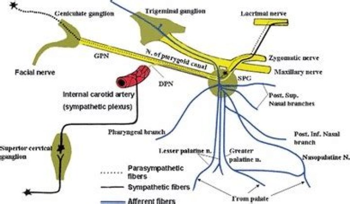
The greater petrosal nerve (or greater superficial petrosal nerve) is a nerve in the skull that branches from the facial nerve; it forms part of a chain of nerves that innervate the lacrimal gland. The preganglionic parasympathetic axons of this nerve synapse in the pterygopalatine ganglion.
What does deep petrosal nerve do?
The deep petrosal nerve carries postganglionic sympathetic axons to the pterygopalatine ganglion, which pass through without synapsing. These axons innervate blood vessels and mucous glands of the head and neck.
Where does the deep petrosal nerve come from?
The deep petrosal nerve is a branch from the internal carotid plexus. The plexus is located on the lateral side of the internal carotid as it courses superiorly. The deep petrosal enters the skull through the carotid canal with the internal carotid artery.
What gives off lesser petrosal nerve?
The lesser petrosal nerve (Figure 26.3) is a continuation of the presynaptic fibers of the tympanic branch of the glossopharyngeal nerve with contributions from the nervus intermedius part of the facial nerve, and the auricular branch (Alderman’s or Arnold’s nerve) of the vagus nerve.Where does the greater petrosal nerve come from?
the greater petrosal nerve, which comes from the facial nerve and runs through the hiatus on the anterior surface of the petrous portion of the temporal bone. a communicating branch with the greater petrosal nerve, which comes from the tympanic cavity, having originated at the glossopharyngeal nerve.
Where is the petrosal sinus?
The superior petrosal sinus is a small, narrow dural venous sinus found within the anterolateral margin of the tentorium cerebelli. It spans from the cavernous to the transverse sinus by coursing through a shallow groove on the superior border of the petrous part of the temporal bone.
What is the hypoglossal?
The hypoglossal nerve enables tongue movement. It controls the hyoglossus, intrinsic, genioglossus and styloglossus muscles. These muscles help you speak, swallow and move substances around in your mouth.
What muscles does facial nerve innervate?
The facial nerve passes through the stylomastoid foramen in the skull and terminates into the zygomatic, buccal, mandibular, and cervical branches. These nerves serve the muscles of facial expression, which include the frontalis, orbicularis oculi, orbicularis oris, buccinator, and platysma muscles.What is nervus intermedius?
The nervus intermedius is the sensory and parasympathetic division of the facial nerve. It contains visceral afferent fibers coming from the taste buds of the anterior two-thirds of the tongue and mucous membranes of the pharynx, nose, and palate.
What is internal carotid nerve?n. A sympathetic nerve extending upward from the superior cervical ganglion along the internal carotid artery, forming the internal carotid plexus.
Article first time published onWhat does the carotid plexus innervate?
Previous research suggest that the external carotid plexus innervates sweat glands on the lateral side of the forehead, whereas the internal carotid plexus innervates the medial forehead’s sweat glands [15]. The external carotid plexus passes filaments to the carotid body and thyroid glands [7].
Where does the greater petrosal nerve exit the temporal bone?
The nerve proceeds anteromedially and exits the superior surface of the temporal bone through the hiatus of the greater petrosal nerve (facial hiatus/hiatus fallopii) and into the middle temporal fossa.
Is lesser Petrosal parasympathetic?
The lesser petrosal nerve (also known as the small superficial petrosal nerve) is the general visceral efferent (GVE) component of the glossopharyngeal nerve (CN IX), carrying parasympathetic preganglionic fibers from the tympanic plexus to the parotid gland.
What are the cranial nerves?
- I. Olfactory nerve.
- II. Optic nerve.
- III. Oculomotor nerve.
- IV. Trochlear nerve.
- V. Trigeminal nerve.
- VI. Abducens nerve.
- VII. Facial nerve.
- VIII. Vestibulocochlear nerve.
What does lacrimal nerve do?
Function. The lacrimal nerve provides sensory innervation to the lacrimal gland, conjunctiva of the lateral upper eyelid and superior fornix, the skin of the lateral forehead, scalp and lateral upper eyelid.
Where is the chorda tympani located?
Anatomical terms of neuroanatomy The chorda tympani is a branch of the facial nerve that originates from the taste buds in the front of the tongue, runs through the middle ear, and carries taste messages to the brain.
What is Jacobson nerve?
Jacobson’s nerve is a tympanic branch of the glossopharyngeal nerve, arising from its inferior ganglion. It enters the middle ear cavity through the inferior tympanic canaliculus, runs in a canal on the cochlear promontory and provides the main sensory innervation to the mucosa of the mesotympanum and Eustachian tube.
What Innervates lacrimal gland?
The sensory innervation to the lacrimal gland is via the lacrimal nerve. This is a branch of the ophthalmic nerve (in turn derived from the trigeminal nerve).
What nerve supplies Stapedius?
The branches of the facial nerve: The nerve to stapedius innervates the stapedius muscle.
What does the Infraorbital nerve innervate?
The infraorbital nerve supplies sensory innervation to the lower eyelid, the side of the nose, and the upper lip (see image below). … The infraorbital nerve supplies sensory branches to the lower eyelid, the side of the nose, and the upper lip.
Are there nerves in the tongue?
General sensation to the anterior two-thirds of the tongue is by innervation from the lingual nerve, a branch of the mandibular branch of the trigeminal nerve (CN V3). The lingual nerve is located deep and medial to the hyoglossus muscle and is associated with the submandibular ganglion.
What is the 11th cranial nerve?
The accessory nerve is a cranial nerve that supplies the sternocleidomastoid and trapezius muscles. It is considered as the eleventh of twelve pairs of cranial nerves, or simply cranial nerve XI, as part of it was formerly believed to originate in the brain.
What is Vestibulocochlear?
The vestibulocochlear nerve (auditory vestibular nerve), known as the eighth cranial nerve, transmits sound and equilibrium (balance) information from the inner ear to the brain.
Is petrosal sinus sampling painful?
Petrosal sinus sampling is an invasive procedure where blood samples are taken from each side of the veins that drain into the pituitary gland. Although the procedure is not painful, you may experience minor pain when the catheter (tubing) is passed through the veins.
What is petrosal sinus?
The superior petrosal sinus is a part of the dural venous sinus system that drains venous blood and cerebrospinal fluid circulating within the cranial cavity. The dural venous sinus system empties into the internal jugular vein and further flows into the cardiovascular circulation via the superior vena cava.
What are brain sinuses?
The dural venous sinuses (also called dural sinuses, cerebral sinuses, or cranial sinuses) are venous channels found between the endosteal and meningeal layers of dura mater in the brain.
What is Stylomastoid foramen?
The stylomastoid foramen is a foramen between the styloid and mastoid processes of the temporal bone of the skull. It is the termination of the facial canal, and transmits the facial nerve, and stylomastoid artery.
Where is 7th cranial nerve located?
Where is the 7th Cranial Nerve located? The two 7th Cranial Nerves (CN VII) are located on either side of the brainstem, at the top of the medulla. They are mixed cranial nerves with BOTH sensory and motor function. CN VII controls the face and is mainly FACE MOVEMENT with some face sensation.
What is geniculate neuralgia?
Geniculate neuralgia is a condition that is caused by a small nerve (the nervus intermedius) being compressed by a blood vessel. Geniculate neuralgia results in severe, deep ear pain which is usually sharp—often described as an “ice pick in the ear”—but may also be dull and burning.
How many nerves are in the face?
SegmentLocationLength, mmLabyrinthine segmentFundus of IAC to facial hiatus3-4Tympanic segmentGeniculate ganglion to pyramidal eminence8-11
Which cranial nerve is for vision?
Optic nerve (CN II) enables vision. Trigeminal nerve (CN V) enables sensation in your face. Vestibular and cochlear nerves (CN VII) enable balance and hearing.